

“What does “fluid buildup in the pericardium” mean?
The outer surface of the heart is covered by a thin membrane consisting of two layers. Between these layers is a lubricating fluid that reduces friction as the heart beats. This fluid is normally present in very small amounts. As a result of certain diseases, this fluid may increase abnormally and accumulate between the layers of the pericardium. The medical term for this condition is pericardial effusion.
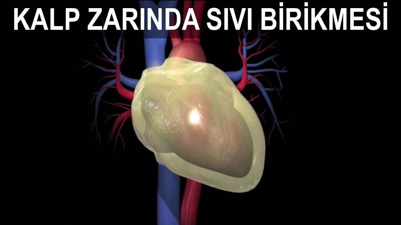
Why does fluid accumulate in the pericardium?
The causes of fluid accumulation in the pericardium (pericardial effusion) are as follows:
- Pericardial infection (viral pericarditis, tuberculosis, etc.)
- Immune system disorders (Lupus, rheumatoid arthritis, etc.)
- Heart attack (bleeding or inflammation)
- Post-heart surgery (bleeding or inflammation)
- Kidney failure (accumulation of toxic substances)
- Cancer (spread of a tumor to the pericardium)
- Hypothyroidism (underactive thyroid gland)
- Accidents and injuries (accumulation of blood)
- Radiotherapy (radiation therapy) to the chest area
- Side effects of certain medications (phenytoin, warfarin, heparin, etc.)
- Other rare causes
What are the symptoms of fluid buildup in the pericardium?
- Easy fatigability (getting tired quickly)
- Chest pain
- Shortness of breath
- Cough
- Fever
- Palpitations
- Swelling in the legs (edema)
- Abdominal swelling (ascites)

How can you tell if there is fluid buildup in the pericardium?
- Blood test
- Chest X-ray
- ECG
- ECHO: Also known as a heart ultrasound. It is the most important test and is essential for a definitive diagnosis.
How is fluid buildup in the pericardium treated?
Medications can be effective if the cause of fluid buildup is inflammation. Nonsteroidal anti-inflammatory drugs (NSAIDs) such as ibuprofen and colchicine are the primary medications. Whether or not there is inflammation, if a large amount of fluid has accumulated or is putting pressure on the heart, this poses a life-threatening risk. In such cases, the fluid must be drained mechanically. Drainage can be performed surgically or non-surgically.
Is surgery or a non-surgical method better for fluid buildup in the pericardium?
The non-surgical method performed with the help of a needle, called pericardiocentesis, is the preferred method. However, not every patient is anatomically suitable for needle drainage. In such cases, surgery is required. During surgery, a portion of the pericardium is removed, and a persistent flow of fluid into the chest cavity is ensured through this opening (window). From there, the fluid enters the bloodstream, then goes to the kidneys, and is excreted through urine. This surgery, called a pericardial window, can also be performed on patients whose fluid accumulates again even after successful needle drainage. Rarely, the entire pericardium may need to be surgically removed (Pericardiectomy). In recent years, the windowing procedure can also be performed non-surgically by inflating a balloon. In the non-surgical method, rather than an incision, usually two balloons are sent through a small entry hole into the pericardium in a deflated state and positioned at the level of the pericardium. When the balloons are inflated, the pericardium is torn, and the resulting opening acts as a window. In this method, called balloon pericardiotomy, unlike surgery, general anesthesia is not required, and no large scar remains. To better understand how the non-surgical method is performed, you can watch the video below.
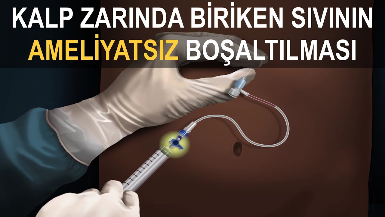

How can fluid accumulated in the pericardium be drained without surgery?
First, using heart ultrasound, namely the ECHO images, we identify the location on the chest where the fluid can be drained most easily. After numbing this area so the patient feels no pain, we advance a needle through the chest wall toward the heart. Once we reach the fluid accumulated in the pericardium, we see that the fluid can be drawn through this hollow needle. Then, we remove the needle and replace it with a thin plastic tube, called a catheter. We drain all the fluid using this catheter. Due to the possibility that the fluid accumulation may continue, the catheter remains between the pericardial layers for a few more days. We remove the catheter on the day we see that fluid is no longer coming out. In most patients, the fluid does not accumulate again. However, in rare cases, fluid may re-accumulate after a few weeks or months. In such instances, it becomes necessary to open a hole larger than a needle hole in the pericardium, which is called a window.
References
- Wang TKM, Klein AL, Cremer PC, et al. An ACC expert consensus statement on the diagnosis and management of pericarditis. J Am Coll Cardiol. 2025 Dec 23;86(25):2691-2719.
- Schulz-Menger J, Collini V, Gröschel J, et al. 2025 ESC Guidelines for the management of myocarditis and pericarditis. Eur Heart J. 2025 Oct 22;46(40):3952-4041.
- Libby P, Bonow RO, and Mann DL, eds. Braunwald’s heart disease: A textbook of cardiovascular medicine, 12th ed. Philadelphia PA: Elsevier; 2022.
- Lasala JM, Rogers JH, eds. Interventional procedures for adult structural heart disease. Philadelphia PA: Elsevier; 2014.