
What is ASD?
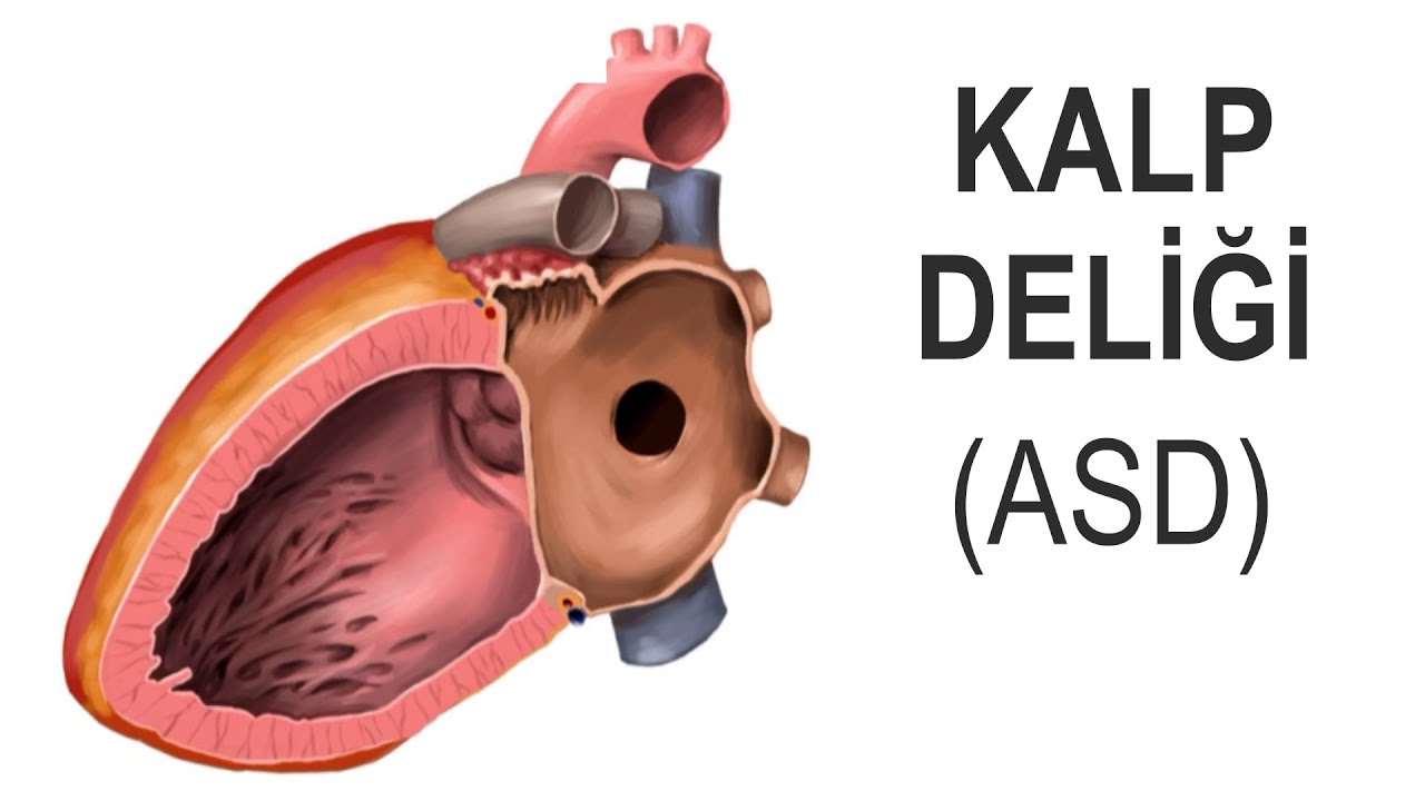
The heart has four chambers. Two chambers are on the left, and two are on the right. The blood in the right chambers contains less oxygen, while the blood in the left chambers contains more. Thanks to the partitions (walls) separating the left and right sides, the blood from the right and left sides does not mix. In some people, developmental issues occur while the heart is forming in the fetus in the womb, and these partitions do not fully develop. Holes remain in the undeveloped sections. One of the most common of these holes occurs in the partition between the right and left atria. In medical terms, this is called an ASD (atrial septal defect). It means “a defect in the wall separating the chambers.”

What are the types of ASD?
ASDs are named based on their location. In 4 out of every 5 cases of ASD, the hole is located exactly in the middle of the septum between the heart’s atria and is referred to as a secundum-type ASD. (It is indicated by the letter B in the figure below.)

How does ASD affect the heart?
The hole, known as an ASD, causes some blood to leak from the left atrium into the right atrium. Because of this leak, the right side of the heart receives more blood than necessary, and its workload increases. Over time, the right side of the heart enlarges due to this additional blood volume. This ultimately impairs the heart’s function. The extent of these effects on the heart and lungs is closely related to the size of the hole; the larger the ASD, the greater the likelihood of causing problems and requiring treatment.
What are the symptoms of ASD?
Although ASD is present from birth, many people do not experience symptoms until adulthood. Sometimes, it may not be detected until the age of 40 or 50. When symptoms do appear, the most common ones are fatigue and shortness of breath. Small holes generally never cause problems because they do not place an excessive workload on the heart. Very large holes, however, can cause symptoms even in infancy; loss of appetite, feeding difficulties, and growth retardation may occur.
What symptoms can ASD cause in a patient?
- Heart failure: Since the right side of the heart is forced to pump a larger volume of blood than normal, it can become tired and weakened over time, eventually leading to the development of heart failure (insufficiency).
- Heart rhythm disturbance: Pathologically enlarged heart tissues may struggle to maintain their electrical balance. Unnecessary impulses may arise from abnormal foci, resulting in an irregular heartbeat.
- Heart valve leakage: When the heart enlarges, the components forming the valves move away from each other, preventing the valves from closing normally. Consequently, blood leakage can occur, especially in the right-sided valve (tricuspid valve).
- Lung problems: The shunted blood passing from the left side to the right side is pumped into the lungs. In the lungs, increased blood flow can cause various issues (pneumonia, etc.).
- Increased blood pressure in the lung artery: This is the high pressure caused by the excess shunted blood passing from the left side to the right side. Its medical name is pulmonary hypertension.
- Stroke (paralysis): Sometimes, if a clot forms in the legs for any reason and this clot breaks loose and reaches the heart through the veins, it can pass through this hole into the left atrium. From there, as the heart pumps blood, it can travel to an artery supplying the brain. It may become lodged in a cerebral artery, blocking blood flow to that area and causing a stroke.
What are the causes (risk factors) of ASD?
The exact cause of ASD is unknown. It may be caused by genetic changes that occur before birth. During pregnancy, factors such as alcohol, smoking, infections like rubella, substance use (e.g., cocaine), diabetes, certain medical conditions, and medications may trigger these genetic issues.
How is ASD diagnosed?
- ECHO (Echocardiography; heart ultrasound): This is the fundamental test for diagnosing ASD. It is a device that works using sound waves. It captures a motion picture of the heart. The hole and the blood leaking through it can be directly seen in most cases.
- TEE (Transesophageal ECHO): In a classic ECHO performed from the chest, the hole in the heart cannot always be seen, but other findings that cause suspicion of its existence may be detected. In this case, performing an ECHO from inside the esophagus, which is adjacent to the heart, provides clearer images. For this, a flexible cable the diameter of a pencil is inserted into the esophagus. At the end of this cable is a special mechanism that allows image acquisition. In this way; the size, shape, and location of the hole are determined in detail. TEE is also used to guide doctors during the non-surgical closure of the hole.
- MRI (MRI; Magnetic resonance imaging): This device takes pictures of the heart using a magnetic field. It is preferred in cases where a diagnosis cannot be made with TEE.
- CT (Computed tomography): This device takes pictures of the heart using X-rays (radiation). This is also preferred in cases where a diagnosis cannot be made with TEE.
Does ASD resolve on its own?
- Almost all ASDs smaller than 5 mm close spontaneously within 2 years.
- 4 out of every 5 cases with an ASD between 5 mm and 10 mm close spontaneously within 2 years. Rarely, they may not close.
- The probability of spontaneous closure for those larger than 10 mm is extremely low.
How is ASD treated?
Even if an ASD does not close on its own, if the hole is very small, it generally does not need to be closed, as it does not cause any problems. However, most ASDs that are not small will eventually require treatment. Treatment can be performed either surgically or using a non-surgical method.
- Surgical closure: This operation is performed by heart surgeons. During surgery, first, a small incision is made in the chest to reach the heart. A patch is preferred to close the hole in the heart. For the patch, a fabric-like material or the patient’s own pericardium (heart lining) can be used. The surgery is performed by stopping the patient’s heart and using a heart-lung pump during this process. Generally, the surgery can be performed through a small incision in the side of the chest. Rarely, robotic surgery may also be preferred.

- Non-surgical closure: This operation is performed by interventional cardiologists or pediatric/congenital cardiologists, not by heart surgeons. Interventional cardiologists are doctors who perform procedures on patients over the age of 18. In the non-surgical method, the hole is closed with an umbrella-like device.


What is ASD closure?
Although “ASD closure” or “ASD closure procedure” refers to the closure of an ASD through either surgical or non-surgical means, in everyday language it is often used to mean “the non-surgical closure of an ASD.”

Prof. Dr. Şükrü Akyüz is an interventional cardiologist. His area of expertise is the treatment of heart diseases with non-surgical methods. He is a proctor (instructor) and consultant for structural heart disease procedures, including ASD closure
Is it better to close an ASD through surgery or using a non-surgical method?
If possible, the non-surgical method is preferred because it is safer. Since there is no scar on the chest, it does not cause aesthetic concerns. However, the non-surgical method can only close ostium secundum-type ASDs, indicated by the letter B in the figure above; for other ASD types (hole types indicated by the letters A, C, D, and E), surgery is the only option*. About 90% of ostium secundum ASD cases are suitable for closure; the remaining 10% require surgery either because the defect is too large or because the anatomical features at the attachment points for the device are insufficient.
(*Note: In recent years, non-surgical closure techniques have also been developed for selected cases of superior sinus venosus-type ASDs (the type denoted by the letter A) where the anatomy is suitable; that is, non-surgical closure may be possible in some cases. However, these techniques are still very new, and long-term outcomes are not yet known. Therefore, surgery remains the first choice for this type of ASD.)
When will ASD be shut down?
If it is observed that the right ventricle of the heart has begun to enlarge due to the effect of blood flowing from left to right, the ASD should be closed. However, in very advanced (neglected) cases, closing the ASD unfortunately does not provide any benefit; it may even cause harm.
What is the success rate of closing an ASD using a non-surgical method?
In general, the technical success rate is greater than 95%.

What are the risks of the ASD closure procedure?
As with any invasive procedure, there are certain risks associated with ASD closure. Serious complications, such as device dislodgement or clot formation on the device, occur in 1 out of every 100 people; death occurs in 1 out of every 1,000 people.
You may be right to be concerned about these risks; but remember: these complications are rare. What matters is whether the procedure is truly necessary. If the decision has been made in accordance with current scientific data and guidelines, avoiding this procedure actually means exposing yourself to far greater risks (such as heart failure) to a much greater extent.
How long does the ASD closure procedure take?
Generally, 1 hour. However, depending on the anatomical characteristics of the cavity, this duration may be shorter or longer.
Is the procedure performed by stopping the heart, as is done in heart surgery?
No. There is no need to stop the heart.
Will I feel any pain during the procedure?
No. The procedure is performed either under sedation or under general anesthesia. The difference is as follows: Under general anesthesia, a tube is inserted into the patient’s airway (intubation) and the patient receives air from a ventilator; under sedation, no breathing tube is inserted, the patient is not connected to a ventilator, and the patient is simply put to sleep while breathing on their own. In both techniques, medications are administered to prevent the patient from feeling pain during the procedure. Performing the procedure under general anesthesia (intubation) is more comfortable for the patient.
When will I be discharged after the procedure?
Most patients are discharged the next day.
Does the ASD closure device come loose?
Although cases in which the implant has reportedly shifted from its original position have been reported in the literature, this is a very rare occurrence. Once the implant has been covered by tissue within a few months, it is no longer possible for it to move.
Can you feel the device in your body?
No.
Will the device remain in the body for life?
Yes. There is no question of removing the device.
Will I need to take blood thinners after the ASD closure procedure?
Yes. Generally, two blood-thinning medications are taken together for the first 2–3 months. One of these is usually low-dose aspirin, and the other is a different medication containing the active ingredient clopidogrel. After 2–3 months, one of these is discontinued; the other is continued until the 6th month. After the 6th month, there is no need to continue taking blood-thinning medications.
What should people with ASD be aware of?
- Most ASD patients have no restrictions on exercising. However, if there is an ASD that needs to be closed, competitive or heavy sports should be avoided until the ASD is closed and for the first month after the procedure.
- After the ASD is closed, it may take up to 6 months for the patch or umbrella-like device to be covered with tissue. During this process, microorganisms entering the bloodstream can directly infect the patch or device via the blood, leading to a serious infection called infective endocarditis. For this reason, for the first 6 months, antibiotics must be taken before bleeding procedures such as tooth extraction or surgery on another organ. Such precautions are not needed after 6 months. However, rarely, a permanent blood leak may continue at the edge of the patch or device; in this case, taking antibiotics before bleeding procedures is always recommended.
- It is often assumed that the metallic structures of the umbrella-like devices, which are made of nickel-titanium alloy, will be affected by the magnetic field of an MRI. However, these devices have weak susceptibility to magnetic fields; therefore, it is safe to undergo an MRI, including 3 Tesla.
- If the ASD is large, lung artery pressure is high, or heart failure is present, going to high altitudes can cause problems.
- In diving, serious problems can occur due to air bubbles in the blood passing through the hole. If diving is to be performed, it is very important to follow diving regulations. In fact, in some cases, such as a large hole, diving should be avoided entirely.
How long does someone with ASD live?
Studies show that if an ASD is closed before it’s too late, life expectancy is close to that of the general population. However, in cases where closure is delayed—that is, when an ASD is closed after the age of 40—life expectancy may sometimes be shorter, though this is not the case for everyone. This is because, by the time the defect is closed, the condition may have progressed over the years to a stage that can no longer be reversed. In other words, right-sided heart failure and high blood pressure in the pulmonary artery may have become permanent.
References
- Gurvitz M, Krieger EV, Fuller S, et al. 2025 ACC/AHA/HRS/ISACHD/SCAI Guideline for the management of adults with congenital heart disease. J Am Coll Cardiol. 2025 Dec 18:S0735-1097(25)07763-0.
- Baumgartner H, De Backer J, Babu-Narayan SV, et al. 2020 ESC Guidelines for the management of adult congenital heart disease. Eur Heart J. 2021 Feb 11;42(6):563-645.
- Behjati-Ardakani M, Golshan M, Akhavan-Karbasi S, et al. The clinical course of patients with atrial septal defects. Iran J Pediatr. 2016 Aug 1;26(4):e4649.
- Radzik D, Davignon A, van Doesburg N, et al. Predictive factors for spontaneous closure of atrial septal defects diagnosed in the first 3 months of life. J Am Coll Cardiol. 1993 Sep;22(3):851-853.
- Libby P, Bonow RO, and Mann DL, eds. Braunwald’s heart disease: A textbook of cardiovascular medicine, 12th ed. Philadelphia PA: Elsevier; 2022.