

What is a virtual angiogram?
Virtual angiography (virtual angiogram) is the imaging of blood vessels using a computed tomography (CT) scanner.
What is a CT angiogram?
CT angiography is synonymous with virtual angiography. CT angiography can be performed on any vessel in the body. However, when we refer to “CT angiography” in this article, we mean the imaging of the heart vessels—namely, “coronary” CT angiography. In CT angiography, it is the X-ray, a type of radiation, that allows us to obtain images. An intravenous dye (contrast agent) enables the vessels, which are normally invisible, to be visualized via X-rays.
What is the difference between CT angiography and conventional angiography?
Both virtual angiography and classic angiography are procedures performed to visualize the heart vessels. In classic angiography, a long and flexible tube (catheter) is sent to the heart through an artery in the groin (leg) or the wrist. Then, dye is injected directly into the heart vessels through the catheter. In virtual angiography, however, no catheter is sent to the heart, and the dye is not injected directly into the heart vessels; it is administered into a vein in the arm or the back of the hand, from where it reaches the heart.
What is the difference between a CT angiogram and a cardiac CT scan?
One of the devices used in the diagnosis of and treatment planning for many conditions—such as coronary arteries, heart valves, the pericardium, and congenital abnormalities—is the computed tomography (CT) scanner. The general term for all of these is “cardiac CT.” Coronary CT angiography is the specific term for the cardiac CT scan performed on the coronary arteries.
Where is a virtual angiogram performed?
Virtual angiography is performed at imaging centers or in the radiology departments of hospitals. A radiology technician performs the procedure under the supervision of a radiologist. The images are then processed using specialized software and interpreted by radiologists. The processed images are also reviewed by cardiologists.
How do you prepare for a virtual angiogram?
You are generally required to fast for 4 hours before the test. However, you may drink plain water up until 2 hours before the test. On the day of the test, you should avoid caffeinated beverages (tea, coffee, etc.), as these can increase your heart rate. Images are not clear at high heart rates, and the reliability of the test decreases. To prevent this, it may be necessary to use a medication called a beta-blocker (Beloc ZOK, Saneloc, Concor, Vasoxen, etc.) to slow your heart rate (more accurately, to bring it back to a normal rate; ensuring it is 50–70 beats per minute). These medications can also be administered directly into a vein if necessary. Additionally, some patients are very anxious, and their heart rates rise significantly due to stress during the scan. In such cases, the temporary use of anti-anxiety medications is quite helpful in controlling heart rate. However, as the number of slices increases, the likelihood of being affected by heart rate decreases, thereby ensuring better image quality. For example, a 1,152-slice device can produce very clear images even if the heart is beating relatively fast. Still, in many patients, clear images can be obtained without a device of such high slice count.

How is a virtual angiogram performed?
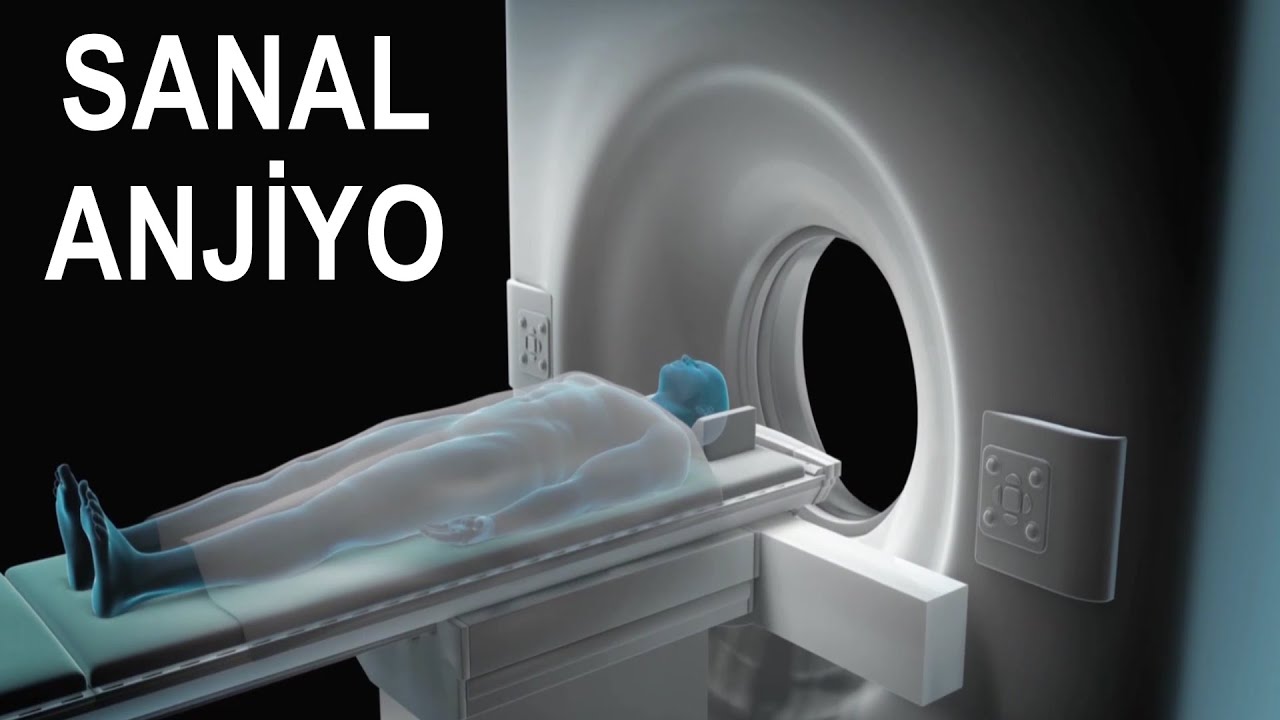
An intravenous line is established in the back of the hand or in the arm. The contrast dye is injected into your body through this line. You will then lie down on a table resembling a stretcher in front of the machine. During the imaging process, you will be asked to remain still and hold your breath periodically to minimize heart movement. When the contrast dye reaches the heart, the machine emits X-rays at that exact moment to capture images of the blood vessels.
How long does a virtual angiogram take?
Although the entire process takes about 15 minutes, the actual active recording time is only 10–15 seconds. In cases involving rapid heart rate, this duration may extend to 1–2 hours.
What are the benefits of virtual angiography?
- Although the image quality is not as high as in classic angiography, it is quite superior. Therefore, in most hospitals today, it has surpassed the stress test (treadmill) for coronary artery disease screening (check-up).
- It demonstrates anatomy and abnormal vascular connections in congenital heart diseases remarkably well.
- Since the imaging process is very brief and does not involve the issues of long duration, noise, or more confined spaces found in MRI, it is a test that patients tolerate much better.
- The risk of complications is much lower than in classic angiography because it does not involve entering the openings of the coronary arteries with a catheter and does not require arterial access to perform the procedure.
Is a virtual angiogram risky?
A virtual angiogram is a safe procedure for most people. However, it does expose the patient to a small amount of radiation. Pregnant women should not undergo a virtual angiogram due to the potential risk of harm to the baby (fetus) in the womb. Additionally, some people may experience an allergic reaction to the contrast dye used. In individuals known to have allergies, a steroid medication (such as cortisol) can be administered several hours before the procedure to reduce the risk of a reaction. Aside from allergies, the contrast dye can rarely cause temporary impairment of kidney function. Therefore, drinking plenty of water before and after the procedure is important for protecting the kidneys. Patients with chronic kidney disease have kidneys that are more sensitive to the contrast agent. It is recommended that these patients be evaluated by a nephrologist before the procedure and receive continuous intravenous fluids starting several hours beforehand.
Does a virtual angiogram provide definitive results?
If the CT angiography result shows “no problems” in the heart vessels, the accuracy rate is near 100%, making it highly reliable. In such cases, there is no need for classic angiography to confirm the findings. However, if a problem is detected, the accuracy rate of the test decreases slightly (80-90% accuracy). The margin of error increases particularly in scans performed without sufficiently lowering the heart rate. In these instances, it may be impossible to distinguish whether a detected narrowing (stenosis) is truly there or if it appears narrowed due to blurred images caused by motion artifacts. To clarify, classic angiography—the test with the highest accuracy—may be required. However, if your cardiologist has reviewed the images personally rather than relying solely on the written report and is confident that the images are clear, the likelihood of an error is quite low. Generally, when CT angiography errs, it is in the form of “overestimating” the degree of stenosis. For example, it may mistakenly show a 30-40% narrowing as 70-80%. The reverse is rare. For this reason, further testing is usually not needed when a narrowing of less than 50% is detected. In some imaging centers, by utilizing an additional feature called CT-FFR, both anatomical and physiological assessments can be performed. That is, if a visual narrowing between 40% and 70% (moderate) is present, the CT-FFR method can determine whether sufficient blood is passing through the stenosis. This significantly increases the accuracy rate of the test. In individuals who have had a stent placed or have undergone bypass surgery, the margin of error is higher compared to other patient groups. In most of these patients, it still provides accurate results. However, stents often create “blooming” (interference) under X-rays, and if the stent diameter is <3 mm, the area inside the stent may not be clearly visible; thus, even if the stent is open, it may appear blocked. Similarly, image quality may sometimes be suboptimal at the connection points (anastomoses) of bypass vessels. In these cases, another imaging method (cardiac scintigraphy or classic angiography) may be necessary. Ultimately, it is important that the center performing the CT angiogram is an experienced facility that frequently conducts coronary CT angiography, that the interpreting radiologist is particularly experienced in cardiac radiology, and that the scan is performed with a modern device of at least 128 slices. A CT angiogram performed and interpreted according to these standards has a much higher accuracy rate compared to the stress test, cardiac scintigraphy (SPECT or PET), and stress ECHO (among these, the test with the highest margin of error is the stress test). For this reason, European (ESC) and American (ACC) cardiology guidelines now recommend CT angiography as the screening test for most patients suspected of having coronary artery blockage.
References
- Virani SS, Newby LK, Arnold SV, et al. AHA/ACC/ACCP/ASPC/NLA/PCNA Guideline for the management of patients with chronic coronary disease. Circulation. 2023 Aug 29;148(9):e9-e119.
- Vrints C, Andreotti F, Koskinas KC, et al. ESC Guidelines for the management of chronic coronary syndromes. Eur Heart J. 2024 Sep 29;45(36):3415-3537.