
What is the aortic valve
The aortic valve is the valve through which blood passes as it is pumped from the heart to the body. It is called the “aortic valve” because it is attached to the aorta, the largest blood vessel in the body. The function of the aortic valve is to allow blood destined for the body to flow into the aorta, but to prevent that blood from flowing back into the heart.
What is aortic valve stenosis?
A problem with the opening of the aortic valve leads to a narrowing of the area through which blood passes, known as the valve orifice. This condition is called “aortic valve stenosis.” It is also commonly referred to as “aortic stenosis” or simply “aortic stenosis.”

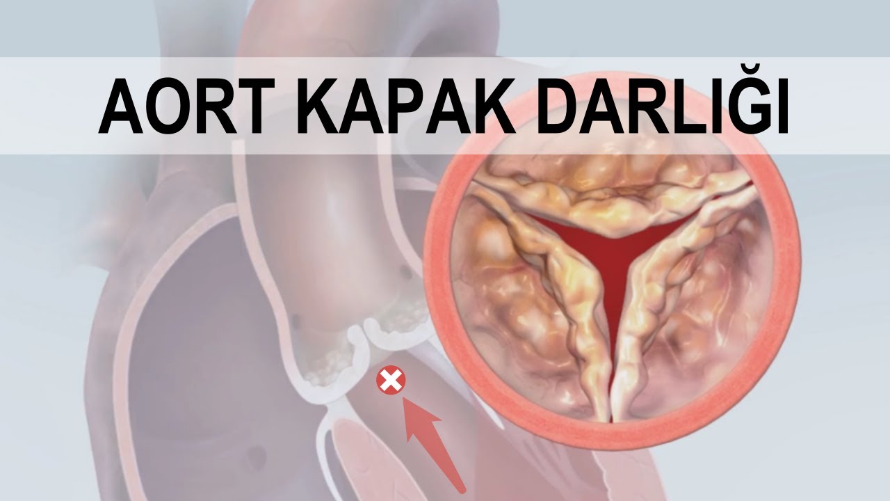
What happens in aortic valve stenosis?
When the aortic valve narrows, blood can manage to pass through this narrowed area in the same volume by increasing its velocity up to a certain point. However, once the stenosis in the valve becomes excessive beyond a certain stage, blood can no longer pass through in sufficient amounts to the post-stenotic area, even if its velocity increases. The blood that cannot pass causes the heart’s main pumping chamber, the left ventricle, to exert more effort; this leads to the thickening (hypertrophy) of the left ventricle and a decrease in its strength. Consequently, over time, the heart wall stiffens and heart failure develops.
What are the symptoms of aortic valve stenosis?
Patients most commonly complain of the following symptoms:
- Getting tired easily (Easy fatigability)
- Shortness of breath (Dyspnea)
- Chest pain (Angina)
- Palpitations
- Dizziness
- Fainting (Syncope)
No complaints are expected in mild or moderate stenosis. For a patient to feel symptoms, the stenosis must reach an advanced (severe) level. The severity of these symptoms can be very mild or very pronounced. Not every patient has to feel all the symptoms listed above; each patient may suffer from a different symptom. Sometimes, some patients with aortic valve stenosis feel no complaints at all, even though the condition is severe, or they may fail to realize and describe them. If severe aortic valve stenosis is left untreated, heart failure will eventually develop, and symptoms and findings related to it will be added to the clinical picture.
What are the causes of aortic valve stenosis?
- Calcification of the valve: As age advances, calcification (degeneration) occurs in the aortic valve. In fact, just as graying of the hair is considered normal with aging, a certain amount of calcification in the valve is also normal as we get older. However, in some people, unfortunately, the calcification is excessive. Sometimes calcification stems from an obvious issue such as chronic kidney failure or uncontrolled hypertension , but in most cases, it is not clearly known why more calcification occurs in certain individuals.
- Congenital abnormalities: The aortic valve normally consists of three leaflets. In some people, however, it is abnormally composed of two leaflets from birth. This is called a “bicuspid aortic valve.” Bicuspid aortic valve is the most common congenital heart disease. It is present in approximately one out of every 100 people. Problems can occur in both the opening and closing of a bicuspid valve. Unlike general calcification, it causes symptoms at relatively younger ages (40s or 50s).
- Damage caused by infections: Microorganisms, especially bacteria, can damage the heart valves either directly (infective endocarditis) or indirectly (rheumatic fever). Aortic valve stenosis due to rheumatic fever develops because pharyngitis or tonsillitis experienced in childhood affects the heart valves due to problems in the immune system. This stenosis appears years later, and these patients almost always have mitral valve stenosis as well.
- Other causes: Chronic kidney failure, autoimmune diseases such as SLE (Lupus), and other rare conditions like radiotherapy (radiation therapy) to the chest area can also cause aortic stenosis.
Who is at risk for aortic valve stenosis?
Aortic valve stenosis is most common in the elderly. In particular, its prevalence increases significantly after the age of 70. Aortic stenosis may also accompany certain other conditions, in which case younger individuals may be affected. In rare cases, infants may be born with congenital aortic valve defects.
How is aortic valve stenosis diagnosed?
- EKG (Electrocardiography): Measures the electrical activity of the heart. This way, rhythm disturbances caused by valve narrowing can be detected and thickening of the heart wall can be indirectly shown.
- ECHO (Echocardiography; cardiac ultrasound): Taking a film of the heart using sound waves. It is the main test in diagnosing aortic valve stenosis. Both the narrowed valve is directly visualized and the severity of the narrowing is calculated by detecting that blood passes through the narrow area more quickly. Sometimes when good images cannot be obtained with classical (surface) ECHO, ECHO (TEE) performed through the esophagus may be needed.
- Chest X-ray: Due to aortic valve stenosis, abnormal enlargement of the heart may be seen. Additionally, the widening of the aortic vessel due to the narrowing can also be detected. Basically, it is useful in distinguishing other diseases such as lung disease.
- CT (Computed Tomography): In some patients where diagnosis with ECHO is difficult, the degree of stenosis can be estimated by looking at the amount of calcification on the valve.
- MRI (Magnetic Resonance Imaging): It is used to detect aortic valve stenosis and its severity when good quality images cannot be obtained on ECHO or when there is discrepancy between ECHO findings and symptoms.
- Cardiac Catheterization: When good quality images cannot be obtained on ECHO or when there is discrepancy between ECHO findings and patient complaints, aortic valve stenosis can be detected by taking measurements with catheters passed through the valve.
- Stress Test (Treadmill): Some patients gradually become so accustomed to the negative effects created by a slowly worsening (narrowing) valve that they unconsciously limit their physical activities in daily life. They may answer the question “Do you have any symptoms?” with “No, I’m doing very well.” However, there may be severe aortic stenosis on ECHO. In this case, doctors may stress the patient’s heart by making them exercise and want to more objectively evaluate whether the patient’s complaints are truly present. In some patients who claim to have no symptoms, stress test results reveal that they actually tire very quickly. Thus, the timing for valve intervention is not missed by mistakenly assuming the patient has no complaints.
How fast does aortic stenosis progress?
If aortic stenosis is mild or moderate, it does not cause symptoms in the patient and no life-threatening problems are encountered. It is sufficient to follow up such patients with ECHO at regular intervals to check whether the stenosis is progressing. In advanced stenosis, treatment is required; because, if left untreated, it carries a life-threatening risk. The rate of progression varies from person to person. In some people, aortic stenosis progresses very slowly and it takes many years for symptoms to appear. In others, this process is very rapid; the stenosis can reach a serious level within a few months.

Can aortic valve stenosis be prevented?
Unfortunately, it cannot be prevented. Although measures such as a healthy diet, lifestyle changes, and medication help protect the coronary arteries, they cannot prevent the progression of aortic valve stenosis. No method has yet been found to reduce calcification.
How is aortic valve stenosis treated?
Since aortic valve stenosis is a mechanical problem, the valve must be replaced with a prosthetic valve through heart surgery or a non-surgical method (TAVI) .

- Heart valve surgery: In this surgery, the narrowed aortic valve is removed and replaced with a prosthetic heart valve. While a full (long) incision is made in the rib cage in classic valve surgery; a half-incision (small incision) is made in new (alternative) methods. Small incisions are also called “minimally invasive methods” or “closed methods” (In fact, these are not “closed” methods; after all, there is an “incision” in the chest, even if it is small). In robotic surgery, only holes are opened in the rib cage rather than an incision. However, in all of them, the heart is stopped in any case. When the heart is stopped, a device called a heart-lung pump performs the task of pumping blood to the body. For more detailed information about heart valve surgery, you can also take a look at our related article.
Belirttiğiniz HTML yapısını, stil özelliklerini ve bağlantıları (linkleri) bozmadan hazırladığım İngilizce çeviri aşağıdadır:
- TAVI (Non-surgical heart valve replacement): In the past, valve replacement could only be performed through surgery. Thanks to advancing technology, the aortic valve can now be replaced through a non-surgical method via the leg (groin) artery. This is called the TAVI method. For more detailed information about the TAVI method, you can also take a look at our related article.

Prof. Dr. Şükrü Akyüz is an interventional cardiologist. His area of expertise is the treatment of heart diseases using non-surgical methods. He is a proctor (instructor) and consultant for structural heart disease procedures, including TAVI.


- Balloon valve expansion: This is a non-surgical method performed using balloon catheters sent to the heart through the leg artery. It is called “balloon valvuloplasty.” It is based on the principle of expanding the narrowed valve area through a tearing effect by inflating a balloon positioned at the level of the valve. No prosthetic valve is implanted. This method is often preferred in children; because if the valve is replaced, the prosthetic valve cannot grow as the child grows, making it too small for the patient in adulthood. Unfortunately, the beneficial effect of the balloon does not last long and needs to be repeated periodically. In adults, it is now performed very rarely because it has been observed that the valve re-narrows very quickly after the balloon procedure; it has been replaced by TAVI. Sometimes it is performed in adults in emergency situations to provide temporary relief for the patient. Ultimately, the definitive treatment is valve replacement.
- Medical (Drug) treatment: Unfortunately, some patients carry a very high risk for heart surgery and are not even suitable for the TAVI method. In this case, medications are used (palliative care). Medications cannot prevent the progression of valve stenosis, but at least they provide relief from symptoms. Other conditions frequently seen in patients with aortic valve stenosis, such as hypertension and heart rhythm disorders, also require the continuation of their own medical treatments. This is because such conditions amplify the negative effects of aortic valve stenosis.
Which is better: a plastic cap or a metal cap?
There are two types of prosthetic heart valves: Mechanical valves and biological valves. Mechanical valves are hinge-like structures (it is more accurate to use the term “mechanical” valve instead of “metal” valve). Since they are prone to blood clots, a strong blood thinner called warfarin must be used for a lifetime . This medication is difficult to use as it frequently interacts with other drugs and certain foods. Mechanical valves are quite durable. They do not wear out easily and function for a very long time; the need for a second replacement is lower compared to biological valves. Therefore, they are often preferred in relatively younger patients (<50 years old). Biological valves, on the other hand, are produced from the pericardium of pigs or cattle. They do not easily form clots; therefore, the use of warfarin is not required. This provides significant comfort to the patient. However, the disadvantage of these valves is that they deteriorate relatively earlier than mechanical valves. For this reason, they are often preferred in older patients (>65 years old) or in cases where warfarin use is risky (e.g., pregnancy). In patients between the ages of 50 and 65, either biological or mechanical valves may be preferred depending on the situation. However, there are many more details than what is written here, and the final choice of valve is made between the doctor and the patient after discussing the pros and cons of both valve types in detail.

What happens to a patient if severe aortic valve stenosis is left untreated?
The only treatment for severe aortic valve stenosis is valve replacement. Medications only help relieve symptoms such as shortness of breath for a limited time; however, the underlying condition continues to worsen.

How long does aortic valve surgery take? How long does TAVI take?
Open-heart surgeries typically last 4 hours, while TAVI procedures take an average of 1 hour. Since clinical and anatomical characteristics can vary from patient to patient, these durations may be shorter or longer.
How long is the recovery period after aortic valve surgery and TAVI?
The recovery process varies depending on the method used. Patients who undergo surgery typically stay in the hospital for a few days, and full recovery takes several weeks. With TAVI, however, patients are usually discharged the day after the procedure, and the recovery period is much shorter.
What are the risks of aortic valve surgery and TAVI?
As with any open-heart surgery, serious risks (complications) may be involved. Most of these risks can also be seen in TAVI. For more detailed information about the risks, you can also visit our heart valve surgery and TAVI sections.
- Blood leakage around the prosthetic valve (paravalvular leak)
- Need for a permanent pacemaker
- Severe bleeding
- Heart rhythm disorders (arrhythmias)
- Infection
- Acute kidney failure
- Heart failure
- Inflammation of the heart lining (pericarditis)
- Stroke
- Death
References
- Otto CM, Nishimura RA, Bonow RO, et al. ACC/AHA Guideline for the management of patients with valvular heart disease. J Am Coll Cardiol. 2021 Feb 2;77(4):e25-e197.
- Vahanian A, Beyersdorf F, Praz F, et al. ESC/EACTS Guidelines for the management of valvular heart disease. Eur Heart J. 2022;43(7):561-632.
- Baron SJ, Magnuson EA, Lu M, et al. Health status after transcatheter versus surgical aortic valve replacement in low-risk patients with aortic stenosis. J Am Coll Cardiol. 2019;74(23):2833-2842.
- Leon MB, Mack MJ, Hahn RT, et al. Outcomes 2 years after transcatheter aortic valve replacement in patients at low surgical risk. J Am Coll Cardiol. 2021;77(9):1149-1161.
- Baron SJ, Ryan MP, Chikermane SG, et al. Long-term risk of reintervention after transcatheter aortic valve replacement. Am Heart J. 2024;267:44-51.
- Otto CM. Timing of aortic valve surgery. Heart. 2000;84(2):211-218.
- Malaisrie SC, McDonald E, Kruse J, et al. Mortality while waiting for aortic valve replacement. Ann Thorac Surg. 2014;98(5):1564-1571.