

What is aortic coarctation?
The aorta is the largest vessel that takes the blood pumped by the heart and distributes it to the body. Aortic coarctation is a congenital narrowing of the aorta. Coarctation means “narrowing” in Latin.
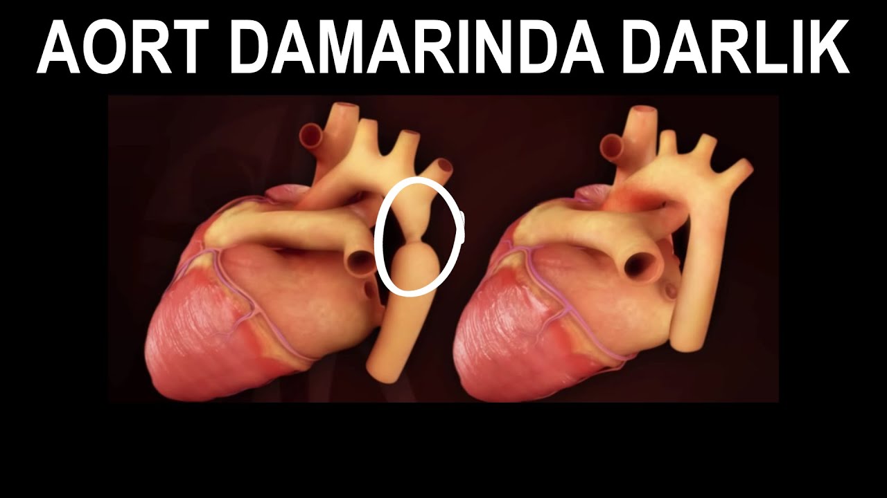

How is aortic coarctation treated?
If aortic stenosis is severe, treatment is either surgery or non-surgical stent placement. Currently, stent placement is the preferred method if possible.


How is a stent placed in aortic coarctation?
First, we take the patient to the catheterization laboratory. We emit X-rays at the level of the aorta. We follow the images obtained this way on the screen in front of us. We perform the procedure by following these images. We numb the groin area. We enter the groin vein with a needle and send a wire through it. Then, we remove the needle and place a flexible sheath (catheter). Through this, we send a wire into the aorta. We advance another long catheter by sliding it over this wire and inject a special dye into the aorta. This dye, called contrast dye, appears black under X-rays, allowing vessels that are normally invisible to be seen. This way, we clarify the location and shape of the narrowing and take measurements. Then, we send the catheter carrying the stent beyond the narrowing. After alignment, we inflate the stent’s balloon. Once the stent is fully expanded, we inject dye again and take control images. After the narrowing is opened, we perform the necessary checks, remove all catheters, and conclude the procedure.

References
- Mazzolai L, Teixido-Tura G, Lanzi S, et al. ESC Guidelines for the management of peripheral arterial and aortic diseases. Eur Heart J. 2024 Sep 29;45(36):3538-3700.
- Isselbacher EM, Preventza O, Hamilton Black J 3rd, et al. ACC/AHA Guideline for the diagnosis and management of aortic disease. Circulation. 2022 Dec 13;146(24):e334-e482.
- Stout KK, Daniels CJ, Aboulhosn JA, et al. AHA/ACC Guideline for the management of adults with congenital heart disease. Circulation. 2019 Apr 2;139(14):e698-e800.
- Baumgartner H, De Backer J, Babu-Narayan SV, et al. ESC Guidelines for the management of adult congenital heart disease. Eur Heart J. 2021 Feb 11;42(6):563-645.
- Libby P, Bonow RO, and Mann DL, eds. Braunwald’s heart disease: A textbook of cardiovascular medicine, 12th ed. Philadelphia PA: Elsevier; 2022.